| Issue |
Eur. Phys. J. Appl. Phys.
Volume 100, 2025
Special Issue on ‘Imaging, Diffraction, and Spectroscopy on the micro/nanoscale (EMC 2024)’, edited by Jakob Birkedal Wagner and Randi Holmestad
|
|
|---|---|---|
| Article Number | 8 | |
| Number of page(s) | 16 | |
| DOI | https://doi.org/10.1051/epjap/2025005 | |
| Published online | 17 March 2025 | |
https://doi.org/10.1051/epjap/2025005
Original Article
Unraveling protein repulsion forces with nanocelluloses: insights from force spectroscopy
Department of Chemistry, Stockholm University, Stockholm SE-10691, Sweden
* e-mail: This email address is being protected from spambots. You need JavaScript enabled to view it.
Received:
17
November
2024
Accepted:
7
February
2025
Published online: 17 March 2025
Abstract
Despite the promising potential of novel surface-functionalized nanocelluloses in advanced technology applications, mechanisms of nanoscaled interacting forces and their origins during interactions between proteins and these materials in biologically relevant environments remain poorly understood. This study explores force interactions between bovine serum albumin (BSA) and three functionalized nanocelluloses, OSO₃⁻-modified CNCs, lignin-residual CNCs (LCNCs), and COO⁻-modified TEMPO-oxidized cellulose nanofibers (TCNFs), to understand protein-nanocellulose interactions using Peakforce quantitative nanomechanical mapping in salt solution at two pH values. The force spectroscopy measured by a protein colloidal probe revealed that TCNFs and LCNCs resist BSA adsorption via pH-dependent repulsion, independent of substrate and protein charge. At pH 3.5, TCNFs showed short-range repulsion (25 nm) against oppositely charged proteins, decreasing to separation distance of 14 nm at pH 7.2. The secondary minima observed at pH 7.2 confirmed the electrostatic-dominant repulsive behavior of TCNFs. LCNCs displayed steric repulsion (13 nm) at pH 3.5 and very long-ranged repulsion (75 nm) at pH 7.2. These repulsion mechanisms, driven by electro-steric repulsion, and hydration forces resulting from formation of a water layer bound to proteins during protein layer compression, deviate from classical DLVO theory. The proposed interaction force mechanisms for protein repulsion were further validated by energy dissipation data and dynamic contact angle experiments. The results showed that TCNFs formed compact layers preserving protein conformation, facilitated by the surface chemical and structural advantages of TEMPO-oxidized TCNFs. While the lignins on LCNCs created flat LCNC-BSA complex layers influenced by lignin chemistry. The findings highlight the importance of optimizing nanocellulose surface chemical properties to enhance protein repellency while maintaining mechanical strength in biological systems.
Key words: Nanocellulose / lignin / PFQNM / force spectroscopy
© J. Li et al., Published by EDP Sciences 2025
 This is an Open Access article distributed under the terms of the Creative Commons Attribution License (http://creativecommons.org/licenses/by/4.0) which permits unrestricted use, distribution, and reproduction in any medium, provided the original work is properly cited.
1 Introduction
Cellulose is a homopolymeric polysaccharide composed of β-D-glucose units linked via β-1,4 linkages, forming a syndiotactic structure with alternating 180° rotations, creating an asymmetric cellobiose repeat unit. The reducing end features a ᴅ-glucopyranose unit in equilibrium with an aldehyde group, enabling site-specific chemical reactions at one end of the chain. Structural advances, such as the natural hierarchical porous structure of networks, strength properties and site-specified activity, have made cellulosic materials very attractive for renewable nanomaterials with desired functions. There are diverse methods for material preparation including acid hydrolysis, oxidation, mechanical method, enzymatic hydrolysis, and hybrid approach [1,2]. Chemical oxidation of cellulose for synthesis of cellulose nanocrystals (CNCs) and cellulose nanofibers (CNFs) is an extensively used process in which the primary −OH groups at position C6 are oxidized to −COOH and secondary −OH groups at positions C2 and C3 to −CHO and −COOH [3–5]. The obtained materials demonstrate a chemical versatility enabled by a wide portfolio of chemical modifications on the hydroxyl groups present at their surface to render tunable surface functionality and environmental friendliness. The isolation processes for nanocelluloses mediated by sulfuric acid, 2,2,6,6-tetramethylpiperidine-1-oxy radical (TEMPO)-mediated oxidation etc. introduces anionic surface functionalities such as −OSO3−, COO− groups on nanocellulose surface. These anionic surface groups is crucial for influencing electrostatic interactions with charged species like metal ions, dyes etc. as adsorbents. One of the most effective methods for introducing functionalized groups such like highly charged COŌ groups onto celluloses is the TEMPO-mediated oxidation. TEMPO-mediated oxidation selectively targets the C6–OH groups on the surfaces of crystalline celluloses. As a result, the C6–OH groups are mostly converted to sodium C6-carboxylate groups during the oxidation of cellulosic surfaces leading to highly ordered distribution of the chemical groups along the cellulose backbones [6–8]. These materials are readily available, renewable, and sustainable and thus they represent a natural resource of incredible importance in today's nanotechnologies [2,9–13]. The motivation for developing lignin-containing cellulose nanomaterials lies in utilizing biowaste or reducing processing with unbleached pulp, while enhancing polymer compatibility, water resistance, and oxidation stability. Understanding of lignin nanoparticle (NP) interactions at small scale during synthesis remains limited [14]. To date, a key challenge in advancing high-tech nanocellulose materials is understanding cellulose–liquid interactions to optimize synthesis routes like designing a single step bio-extraction procedure and surface functionalization methods, enabling tailored functionalities for emerging applications in coating technique, biosensing and drug delivery [15–17]. Lignin's inherent structural and surface chemical complexity, coupled with the diversity of liquid–liquid interactions and lignin types necessitates further in-depth research at very small scales to fully harness its potential by fine-tune its surface properties. This demands integration of colloid and interface science with nanocellulose technology, as critical material properties are strongly influenced by these interactions. Addressing this complexity demands interdisciplinary in situ approaches to study the intricate surface chemical mechanisms during interactions with molecules.
In situ atomic force microscopy (AFM) are proven powerful tools to measure localized surface chemical information in real time, providing insights into fundamental mechanisms of force interactions at nanoscale between any surfaces of aforementioned nanomaterials [18–20]. Force spectroscopic studies using AFM for protein-repelling properties by conventional materials dextran, poly(ethylene oxide), and polystyrene indicated that resulting forces depend on various surface properties, including side chain structure and charge density [21–24]. Force spectroscopy studies for bovine serum albumin (BSA) −lignin interactions showed that protein adsorption is driven by electrostatic, hydrophobic, and van der Waals forces, dependent on ionic strength and protein composition. Proline residues promote protein corona formation on lignin nanoparticles, but their role in intermolecular interactions remains underexplored [25]. We previously showed that newly developed CNC, TCNFs and LCNCs exhibit superior antifouling performance for protein repelling and controlling Escherichia coli (E. coli) cell growth. We further developed in situ Peakforce quantitative nanomechanical mapping (PFQNM) techniques to investigate the nanomechanical properties of E. coli during its in situ growth on nanocellulose films under operational conditions [26–31]. Force spectroscopy using colloidal AFM probe techniques has been extensively utilized to quantify interaction forces and their origins between biological macromolecules and cellulosic systems [32–35]. The advantages of using colloidal probe techniques with spherical particles of defined radius include: 1) the ability to analyse forces more quantitatively using classic intermolecular force interacting model e.g. Deryaguin–Landau–Verwey–Overbeek (DLVO) theory; 2) higher total force, allowing for more sensitive measurements. 3) The versatility to create various probes by attaching particles of different chemical compositions to cantilevers; 4) the ability to measure hydrodynamic forces [36]. Very recently, we developed novel TCNF nanocellulose colloidal probes to further investigate the force interactions between TCNF and BSA under in situ conditions at pH 3.92, near the isoelectric point (IEP) of BSA [37]. Despite recent progress, a significant knowledge gap remains in understanding interaction mechanisms between cellulose nanomaterials and biomolecules (e.g., proteins and living cells) at solid–liquid and liquid–liquid interfaces. The development of larger surfaced colloidal probes modified with biomolecules for precise force measurements is still underexplored [5]. Closing this gap is essential for advancing colloidal science and controlling non-specific adsorption at these interfaces, and for new era of biology the mechanobiology, which is study of how cells generate, sense, and respond to mechanical forces, shaping the future of cellular form and function [38,39].
The motivation for using three types of nanocellulose—lignin-residual cellulose nanocrystals (LCNC), OSO₃⁻ cellulose nanocrystals (CNC), and TEMPO-oxidized cellulose nanofibrils (TCNF)—in a comparative study of force interactions lies in their chemical modifications. These nanocelluloses were functionalized similarly, resulting in distinct functional groups and structural properties that significantly influence their interaction behaviors. LCNC retains lignin, an aromatic polymer with hydrophobic functionalities, antioxidant and antimicrobial properties, Lignin's inherent surface chemical complexity renders diversity of liquid–liquid force interactions. TCNF, with uniformly distributed carboxyl and hydroxyl groups on CNF, is highly charged, hydrophilic, and flexible, offering a large surface area. CNC, derived via sulfuric acid hydrolysis, features rod-like particles with high crystallinity, sulfate groups making it negatively charged and hydrophilic. The surface chemistries (lignin in LCNC, sulfate in CNC, and COO⁻ in TCNF) and physical properties of these nanocelluloses significantly influence interaction and binding forces between molecules or materials, introducing unique interaction behaviors. The motivation for this study stems from the functional and structural variations of these materials, which enable diverse and in-depth exploration of interaction forces in colloidal charged liquid phases. By comparing these nanocelluloses, this work elucidates how structural-surface chemistry relationships govern interactions, paving the way for their application in advanced technologies.
This study firstly aims to enhance the accuracy of force measurements using a newly developed BSA-functionalized colloidal probe, which offers a larger surface area for measuring total forces with higher precision compared to nanofiber-coated AFM tips. Additionally, it addresses the limited understanding of pH-dependent interfacial forces between proteins and TEMPO-oxidized nanocelluloses or lignin nanomaterials in biologically relevant environments. To clarify these mechanisms, nanoscale interacting force and their origins between BSA and surface-functionalized nanocelluloses (OSO₃⁻-CNCs, lignin-containing CNCs, and COO⁻-CNF) were quantified using force spectroscopy, PFQNM and dynamic contact angle measured in biologically relevant salt electrolyte at varying pH levels. This study provides insights into the influence of surface chemistry on protein adsorption, contributing to the understanding of protein-nanocellulose interactions in biological and surface colloidal contexts.
2 Material and methods
2.1 Materials
The characterized materials were acquired using the methods reported in our previous studies [28,40]. In short, O-SO3− modified CNC (CNCs) suspensions (5 wt. % in water, crystallinity degree 76.3 ± 5.6%) were purchased from CelluForce, Canada. The CNCs were isolated using sulphuric acid hydrolysis and carried anionic charge on the surface. Lignin residues grafted CNC (LCNCs) were synthesized in our lab (1.4 wt.%, crystallinity degree 64.8 ± 2.0%) [41]. LCNCs were prepared using partial bleaching for the residual production of bioethanol from wood containing 50% (w/w) cellulose, 42% (w/w) lignin, and 8% (w/w) extractives using high-pressure homogenization. TEMPO mediated COO− modified CNFs (TCNFs) (1.1 mmol/g carboxyl groups, 2.0 wt.%, crystallinity degree 60%) were produced by surface carboxylation of CNFs using TEMPO as the catalyst and subsequent mechanical disintegration. TCNF surfaces were partially carboxylated and hence negatively charged. All three nanocellulose materials possess a high surface-to-volume ratio and high surface charge density with negative charges in a wide pH range. Their surfaces are intrinsically hydrophilic owing to the presence of hydroxyl and carboxyl groups [42]. Structural details of the chemical groups grafted on the CNC, LCNC and TCNF confirmed by Solid-state 13C Nuclear Magnetic Resonance (NMR) spectra were reported elsewhere [28,37].
2.2 Zeta potential measurements
The surface charges of the nanocellulose dispersions were determined using Zeta potential measurements performed with a Zetasizer Nano ZS (Malvern Panalytical Ltd.). Three 5 mL samples of the nanocellulose and protein suspensions in phosphate-buffered solutions (PBS) solutions, with nanocellulose concentrations matching those used for preparing AFM samples on mica, were prepared. Portions of these samples were transferred into DTS0012 cuvettes, into which a universal dip cell kit (ZEN1002) was carefully placed. Each sample underwent five measurements, with each measurement consisting of 10 repeated cycles. The standard deviation calculated from the five measurements was recorded as the error for each sample.
2.3 Scanning electron microscopy − energy dispersive X-ray spectroscopy (SEM-EDS)
Naked AFM probes, and probes functionalized with protein before and after PFQNM measurements, were analyzed using scanning electron microscopy (SEM) on a JEOL JSM-7000F microscope (Japan). The probes were mounted on conductive carbon tape without any additional coating to preserve the integrity of the protein layers. SEM imaging was conducted at 5 kV, with a 15 mm working distance used for EDS measurements. Post-experiment SEM imaging was performed on the AFM probes to inspect for defects or evidence of contamination.
2.4 Colloidal probe functionalization and PFQNM measurements
BSA lyophilized powder ≥96% with a molecular weight (Mw) of 66 kDa (40 × 140 Å) were purchased from Sigma Aldrich. The BSA functionalized probe was made by referring the method in reference [43]. In brief, the cantilever (ScanAsyst-fluid probe, Bruker) was cleaned in acetone for 5 minutes followed by 20 minutes of ozone irradiation. Subsequently, the cantilever was immersed in a 50 µl drop of BSA (0.5 mg/ml in 0.1 M sodium bicarbonate, pH 8.3) overnight at 37°C in a humidified incubator. To eliminate unbound protein, the cantilever underwent three washes in PBS (pH 7.2), followed by fixation of the BSA using a 1.0 wt.% glutaraldehyde solution for one minute. Prior to use, the cantilever was washed three times in PBS. This method offers several advantages including the system has been well-characterized and exhibits high affinity. We aimed to avoid the use of pre-deposited (3-aminopropyl) triethoxysilane (APTES) to minimize unwanted forces originating from saline monolayers during force spectroscopy measurements in a salt electrolyte. The morphology of the BSA probe is depicted in Figure 1. The scanning electron microscope (SEM) image demonstrates the uniform and firm distribution of BSA layers on the BSA modified AFM tip (i.e. BSA probe). The thickness of the protein monolayer is approximately 200 nm, as shown in the inserted SEM image. These layers remained intact after PFQNM experiment (see SI). Additionally, the AFM image confirmed that the size of a single protein nanoparticle (NP) is also around 200 nm. Thus, the protein layer at the apex of the colloidal probe corresponds to a single BSA nanoparticle. The SEM images of a BSA-functionalized probe in SI, taken after PFQNM measurements on nanocellulose samples in electrolytes, demonstrate that the protein coatings remained intact without any visible defects or cracks. Elemental distribution on the protein coatings, as revealed by EDS profiles, is provided in the SI.
PFQNM experiments were conducted using a Bruker Dimension Fast-scan instrument. The Derjaguin-Muller-Toporov (DMT) model, which considers adhesion beyond the contact area, was employed for calculating the elastic modulus. This approximation is considered safe in our scenario due to the small indentations, weak adhesion in aqueous environments, and an acceptable difference of approximately one order of magnitude in the curvature between the cellulose surface and the AFM tip [36,44]. All in situ PFQNM experiments were conducted in phosphate-buffered saline (PBS) electrolytes with a constant salt concentration of 150 mM, at two pH values: neutral (pH 7.2) and acidic (pH 3.5). PBS was selected to simulate a biologically relevant environment. Pre-prepared PBS at pH 7.2 (150 mM) was purchased from Sigma, while PBS at pH 3.5 was freshly prepared using deionized water (Milli-Q System). The pH was adjusted to 3.5 using acetate and to 7.2 using phosphate, ensuring a constant ionic strength of 150 mM. All chemicals used were of analytical grade. Each sample was measured at both pH 3.5 and 7.2 without drying the samples between measurements. The data presented here reflects measurements on samples that were never dried. For each material, separate samples were measured: one at pH 3.5 and another at pH 7.2. Details for data analysis, fitting procedure for PFQNM data and force distance curve analysis can be found in SI.
 |
Fig 1 A representative (left) SEM photograph the BSA coated AFM tip comparing with the SEM image of a naked probe, and (middle) an AFM image of a single BSA nanoparticle (NP) dropped on a mica surface. (Right) Representative SEM image of the protein probe marked with the size of the apex area of the tip. |
2.5 Dynamic contact-angle analysis (DCA) by Wilhelmy plate method
The freshly cleaved mica substrate was coated with suspensions of BSA, CNC, TCNF, and LCNF in PBS solutions at pH 7.2 and pH 3.5, and fastened to a microbalance with a clip using a force tensionmeter (K100, KRÜSS, Germany). A vessel containing 40 ml of either PBS as control or 5 mg/ml BSA PBS solution at the two pH values was then moved towards the substrate, initiating the measurement upon first contact, defined as an immersion depth of 0. The sample was then dipped at a velocity of 1.0 mm/min to an immersion depth of 10 mm and retracted to 0 mm. One cycle was recorded for each coating on mica, taking 20 minutes per experiment. Experiments were conducted using both phosphate-buffered solutions (control) and phosphate-buffered BSA protein solutions (5 mg/ml). The values of θAdv and θRec for hysteresis calculation were the position-dependent averages of θ when equilibrium was established during the descending and ascending positioning using Wilhelmy plate method [45,46]. In thermodynamic equilibrium, the contact angle is related by Young's equation to the interfacial energies. When for example placing a drop on a surface its contact angle can assume any value between the advancing θAdv and receding θRec contact angles, depending on how the drop is placed. The difference between the advancing (θAdv) and receding (θRec) contact angles, known as hysteresis (Δθ = θAdv − θRec), which is caused by the nano scopic structure of the surfaces indicating surface homogeneity [47]. Hysteresis is influenced by the distribution of surface areas acting as barriers. A more homogeneous surface results in smaller hysteresis. In dynamic contact angle measurements, the θAdv is more sensitive to changes in high contact angle regions if most of the surface has low contact angles, while the θRec is more sensitive to changes in lower contact angle regions due to energy dissipation relating to surface roughness [48–50]. The principle of dynamic contact-angle analysis (DCA) can be found in SI: S (9∼12). 2D Fast Fourier Transform (FFT) was used for studying spectral frequencies present in AFM images of BSA NP coated nanocelluloses after DCA measurements, the principle can be found in S13.
3 Results
The isoelectric point (IEP) of BSA is 4.7, the BSA is net charge positive below IEP, and net negative above IEP [23]. This is proved by Zeta potential data in Figure 2. The Zeta potential values measured for the three nanocellulsoe matrials are all negative at each of the pH values. Protein layers are positively charged when the pH is below their IEP (3.5) and negatively charged when the pH is above IEP (7.2). This is also agreed with reference [51]. The surface charges of the BSA layers coated on probe is positive 11.8 mV ± 1.12 mV at pH 3.5 (below the IEP). Whereas at pH7.2 (above the IEP) the protein layers are negative and zeta potential is −6.7 mV± 1.84. As illustrated in Figure 2, the structures of the surface chemical modified nanocelluloses are composed of bounding chemical groups of O-SO3−, COO− or lignin residues owing groups of COO, CHO− and phenolic C10H12O3 on the nanocellulose backbones. The surface potential data has the relation with the measured force according to linear Poisson-Boltzmann equation [52].
 |
Fig 2 Schematic illustration of the structures of the functional chemcail groups OSO3 (H+) on CNCs, COO-(H+) on TCNFs and lignin residues on CNCs. Exact protonated or deprotonated form of the group will depend on the pH condition [42]. Zeta potential results depicting three nanocellulose and BSA (0.5 mg\ml) suspension samples obtained in PBS buffer solutions at pH 3.5 and 7.2, respectively. |
3.1 PFQNM maps of the nanocelluloses measured by BSA colloidal probe
Figures 3–5 show sequential in situ PFQNM mappings of morphology (Z-height), elastic modulus, and adhesion force mapping of the CNC, TCNF and LCNC samples obtained after 180 min (3 h) of immersion in PBS electrolytes at either of the two pH conditions. Table 1 listed the fitted values of the height, elastic modulus and adhesion force for the mappings of PFQNM for each of the material respectively.
At both pH levels, TCNFs demonstrated the highest elastic modulus and adhesion force, followed by LCNCs and CNCs. This can be attributed to ion adsorption driven by the high surface charge density of TCNFs. The abundance of surface charges on TCNFs likely increased fiber interactions and facilitated crosslinking in the presence of salt ions [53,54]. The PFQNM results showed a consistent trend in elastic modulus data, in line with previous findings by us using a bare probe [42]. Notably, the difference in adhesion forces between TCNFs and LCNCs at the two pH values was minimal, with LCNCs exhibiting a very small adhesion force. The high elastic modulus values observed for TCNFs and LCNCs at pH 7.2 were primarily attributed to salt adsorption during water uptake [55]. This further highlights how the surface charges and unique structural properties of TCNFs and LCNCs contributed to their superior nanomechanical performance and enhanced ion adsorption, particularly when compared to pristine CNCs.
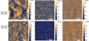 |
Fig 3 in situ PFQNM images of CNCs obtained in in PBS buffer after 3 hours of immersion at pH 3.5 (upper panel) and pH 7.2 (bottom panel) (scale bar: 2 μm). |
 |
Fig 4 in situ PFQNM images of LCNC obtained in PBS buffer after 3 hours of immersion at pH 3.5 (upper panel) and pH 7.2 (bottom panel) (scale bar: 2 μm). |
 |
Fig 5 in situ PFQNM images of TCNFs obtained in PBS buffer after 3 hours of immersion at pH 3.5 (upper panel) and pH 7.2 (bottom panel) (scale bar: 2 μm). |
Peak values of the Gaussian fitted distribution of Z-height, elastic modulus and adhesion of the PFQNM measured for the three materials at pH 3.5 and 7.2 respectively.
3.2 The force interaction measured for nanocelluloses during BSA compression
The force distance (FD) profiles depicted in Figure 6 delineate the distinctions observed in terms of the range, steepness, and magnitude of force interactions at the deprotonated (pH 7.2) and protonated (pH 3.5) chemical groups for each type of nanocellulose. The pH- and charge-dependence exhibited across all cases is closely associated with the surface charge density, structural layers of the nanocellulose, and the structural alterations in BSA conformation [56–59]. The following discussions on the force-distance (FD) data are grounded in this premise and are compared with the results from our previous work [42]. The force measurements for protein interactions with CNCs, LCNCs, and TCNFs across different pH conditions reveal critical insights into their repulsion and attraction behaviors, which are dependent on surface charge and separation distance. For CNCs, as shown in Figure 6a, during tip compression, the repulsion force at pH 3.5 was stronger than that at pH 7.2, despite the repulsion occurring at a similar separation distance of approximately 23 nm for both pH levels. The steepness of the repulsion, reflected in the log(F) plot, was at a comparable level for both conditions.
In the case of LCNCs, as shown in Figure 6b, the repulsion force at pH 7.2 was much stronger than at pH 3.5. Also, the repulsion occurred over a much longer separation distance at pH 7.2 (around 75 nm), compared to approximately short range of 25 nm at pH 3.5. The repulsion force decayed much more slowly at pH 7.2 than at pH 3.5, indicating a steric dominant repulsion, possibly due to the large molecular weight of lignin groups. For TCNFs, as depicted in Figure 6c, the repulsion force during tip compression was much shorter-ranged, occurring at a separation distance of around 13 nm, significantly shorter than that for CNCs and LCNCs. The increase in pH from 3.5 to 7.2 resulted in a substantial rise in both the magnitude and steepness of the repulsion force. The secondary minima measured at pH 3.5 was likely due to the temporary electrostatic attraction between the positively charged BSA and the protonated TCNF surface [42]. When two polymer-coated surfaces approach each other the entropy of confining the polymer molecules results in a repulsive force, known as the effect of “steric” or “overlap” repulsion. This happens when concentration of segments in the gap increases, which is another contribution to the increased osmotic pressure. However, the weak overlap approximation for the steric force is less general than that for the double layer force since it relies on assumptions that are not always valid in force field theory [60]. The gradient of the steric force between a sphere and a planar surface is calculated according to weak overlap approximation by assumptions. First, the segment density in the dilute tail region often decays exponentially with distance from the surface. The second assumption is that the segment density is so small that the conformation of the polymer layer is not affected by the presence of a second polymer layer in its close proximity. This assumption can only be reasonable at large separations where the segment density is low [61]. In this study, the logarithmic scaled force profiles for TCNC and LCNC showed this effect at long range of separations. An exponentially decaying steric force was detected at large separations (ca. 2–20 nm) for interactions between BSA-LCNC and BSA-TCNF. Most interestingly, the logarithmic scaled force profiles for LCNCs at pH 3.5 clearly showed the significant predominance steric effect for the interaction of oppositely charged surfaces. The measured repulsion force at shorter separations became steeper. In addition, the exponential decay observed in these materials is much more pronounced than that measured for the same nanocellulose types when using a bare probe, when compare results of force profile in our previous studies [42]. This supports the conclusion in this work that the repulsion forces for and LCNCs at very short distances arise from steric interactions. The Debye length of our system is only about 0.8 nm. Given that the measured forces at tip compression are always repulsive between two surfaces at each pH condition, and decays exponentially with separation at the larged separations. These deviations are caused not by break down of DLVO rather by the presence of additional forces such as steric or hydration forces [33,36,58,59,62–65]. For all three cases, the force decays at larger separation distances (>20 nm) are not exponential, indicating the dominance of double layer electrostatic force at these longer ranges [59]. These findings demonstrate that the interactions between BSA and nanocellulose surfaces are highly dependent on pH and the surface properties of the nanocelluloses, with TCNFs exhibiting the strongest repulsive forces across both pH conditions. During tip retraction, attraction force was measured for all three materials at pH 3.5, likely due to the electrostatic attraction between the oppositely charged surfaces. The adhesion force profiles measured at tip retractions are related to the adhesion force results of PFQNM in Section 2.1. Due to the very short Debye length in our system, there may be an uncertainty of a few nanometers when defining the zero-distance, especially for soft surfaces such as the adsorbed BSA layers. This uncertainty arises because, on soft or deformable surfaces, such as those formed by protein adsorption, the exact point of contact is difficult to pinpoint with precision.
Figures 7a and 7b presents a comparison of the force response during the tip compression only for each case at the pH conditions. In Figure 7a, at pH 3.5, the features in the force profile for three materials are noteworthy. Notably, there is an absence of jump-in attributed to attractive primary minima forces before short-range of separation [62]. The interaction between the oppositely charged surface of BSA and the surface of nanocellulose is primarily dictated by repulsion. This electrostatic repulsion corresponds to the overlap of electrostatic double layer forces originating from the BSA layers [56]. The repulsion forces were measured at the greatest separation distance for CNC (29 nm), followed by TCNF (25 nm), and LCNC (13 nm). This range of distances is related to the surface charge density of the three materials. The surfaces of protein layers and nanoculluloses are similarly charged at pH 7.2. In Figure 7b, the much shorter separation distances (14 nm) in TCNF reveals that electrical double layer force dominanted force interaction. Notably, the repulsion force for TCNFs was one order of magnitude stronger than that of LCNCs, due to high surface charges. A noticeable secondary minima attraction measured for TCNFs is observed at separation of 22 nm, indicating the dominance of electrostatic forces that surpass the double layer repulsion, attributing to the electrostatic screening on the surface charge of TCNFs [62].
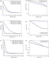 |
Fig 6 Representative force-distance approach and retraction curves (a, b, c) and logged (force) vs. distance at tip approach (a', b', c') measured between BSA tip and (a, a') CNC; (b, b') LCNC; (c, c') TCNF obtained in PBS at pH 3.5 and 7.2 respectively. |
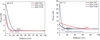 |
Fig 7 Comparison of the representative FD curves at tip approach recorded between BSA and the three materials (a) pH 3.5, and (b) pH 7.2. |
3.3 Energy dissipation of the nanocelluloses during BSA compression
Figure 8 displays the fitted dissipation values acquired at the peak position from the Gaussian fitting of the PFQNM dissipation map for each material. The dissipation measured during the interaction between BSA layers and TCNFs is 348.2 ± 0.9 eV, surpassing that observed for CNCs or LCNCs. We propose that when chemical groups protrude extensively into the electrolyte solution, their confinement into a smaller space during compression by BSA layers leads to unfavourable entropy of confinement, causing the dissipation [62].
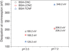 |
Fig. 8 Fitted dissipation energy at tip compression as a function of pH for each of the material. |
3.4 Reduction of protein adsorption by nanocelluloses using dynamic contact-angle hysteresis analysis (DCA)
The change in hysteresis can occur when the surface structure is reorganized and changes occur through, e.g., adsorption processes of BSA [45]. We calculated the Δθ for each of the materials as shown in Figure 9, Table 2, and SI Figure S11 and Table S12. The measured hysteresis (Δθ) is smallest for TCNF, followed by LCNC and CNC. The hysteresis for each material is smaller at pH 7.2 than at pH 3.5. The differences in Δθ measured for TCNF, CNC, and LCNC at pH 3.5 are significant, which is due to the oppositely charged surfaces of BSA and the nanocelluloses. The contact angle hysteresis force (FCAH) is calculated using equation 1,
 (1)
(1)
in which γLV (mN/m) is liquid-vapor interfacial tension. θAdv and θRec are the advancing and receding contact angles, respectively, to quantify the maximum pinning force (FCAH) that the solid coating substrate could provide [66–68]. The measured hysteresis (Δθ) and (FCAH for surfaces of TCNF, LCNC and CNC, and of BSA on mica as a control during protein adsorption is shown in Figure 9 and Table 2.
The results demonstrated two key points: first, negatively charged nanocelluloses reduce the adsorption of similarly charged proteins at pH 7.2, compared to their repelling properties against protein adsorption when the BSAs are positively charged at pH 3.5. Second, the lowest Δθ measured for TCNFs at both pH levels indicates that TCNFs possess super liquid-repellent surfaces, which effectively prevent the formation of a BSA monolayer compared to the nanocrystals. TCNF exhibits the best capacity to reduce BSA adsorption regardless of pH conditions. The low contact angle hysteresis, and super liquid-repellent surfaces of TCNF result from the high mobility of the main chain of COŌ groups tethered to TCNFs. Consequently, the surfaces of TCNF exhibit a liquid-like lubricating effect on droplets, resulting in low contact angle wetting hysteresis for water. The water droplet was in stick-slip motion during wetting, i.e.: intermittent contact line pinning causes the water droplet to switch between sticking and slipping during protein adsorption. The hysteresis measured for LCNC in BSA PBS solution at pH 3.5 is much lower than that measured in PBS alone in contrast to that for TCNF and CNC, suggesting the forming of uniform protein layers on LCNC in pH3.5. (See SI-Fig. S12(A)) The hysteresis measured for CNC in BSA PBS solution at pH 3.5 is significantly higher than that in PBS alone (see Fig. S11). the adsorption of BSA resulted in heterogeneous protein layers on CNC in BSA solution at pH 3.5, while the BSA layers were likely uniform in the BSA solution at pH 7.2, as evidenced by the lower hysteresis measured for CNC at pH 7.2.
As shown in Figure 9b, the low pinning force measured for TCNF can be attributed to the highly ordered distribution of COŌ groups and the strong polarization of the COŌ head groups on TCNFs. This results in a dominant slipping motion, which surpasses the slipping motion seen on CNC and LCNC surfaces. Consequently, similarly charged BSA molecules are effectively repelled [46,47,68]. In contrast, the higher pinning force observed for LCNC at pH 7.2 indicates BSA adsorption in the early stages, likely due to the less organized structure and lower surface charge of LCNC nanocrystals. Interestingly, at pH 3.5, the pinning force for LCNC is weaker compared to TCNF, likely due to the influence of lignin's natural hydrophobicity, which promoted slipping despite the oppositely charged surfaces. The results suggest that the water droplets exhibited a slipping motion, reducing the interaction strength. CNC, on the other hand, displayed the strongest pinning force for BSA adsorption under both pH conditions. These results align with the mechanism of protein repulsion observed in force spectroscopy (Fig. 7), where the structural and surface chemistry advantages of TCNF and LCNC come into play. At pH 3.5, the significant long-range steric repulsion observed for LCNC is likely due to the rapid formation of protein monolayers on the oppositely charged lignin surface, which help maintain the integrity of BSA. This interaction makes the lignin groups more hydrophilic, preventing further protein adsorption. In contrast, for TCNF at pH 7.2, the sharp increase in short-range electro-steric repulsion upon BSA compression is attributed to the liquid-like, slippery nature of the TCNF surface, which preserves the BSA structure during compression. In both cases, the formation of water layers likely contributed to the repelling properties [62]. In contrast, CNC exhibited the strongest pinning force for protein adsorption in BSA solutions across both pH conditions. In addition, this conclusion is approved by the AFM imaging result in Table 2 and Figure S12(A), the roughness of BSA coating on TCNF is smallest, follows by LCNC and CNC. During two-dimensional Fast Fourier transform (2D FFT) analysis, filtering is achieved by selecting the intense spots, and removing the rest of the image in Fourier space. Carrying out an inverse FFT gives the image on the right the selected features are preserved and the noise is removed. The BSA monolayer adsorbed TCNF showed the best uniformity, as that is evidenced by the lowest frequency noise in the inversed FFT image. BSA monolayer covered all over the LCNC, big BSA clusters formed on CNC. The AFM results supported the mechanism of protein adsorption revealed by results of DCA. The AFM images of BSA nanoparticles on nanocelluloses after DCA at pH 3.5 did not show significant differences, likely due to the opposite surface charges of the proteins and nanocelluloses (see SI-Fig. S12(B)).
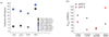 |
Fig. 9 (a) Dependence of measured average hysteresis (Δθ), and (b) pinning force (FCAH) on pH for CNC, LCNC, and TCNF coatings during protein adsorption in PBS-buffered solution containing 5 mg/ml BSA*. |
Roughness, Z-height of the AFM images of nanocelluloses after DCA measurements in electrolyte pH7.2. The pinning force (FCAH) measured for the nanocelluloses during in situ protein adsorption in BSA (5 mg/ml) in PBS solution at the two pH values.
4 Discussion
4.1 Fore profile revealed the non-specific repulsion by surface chemistry of TCNF and LCNC driven protein formation
The force spectroscopy profiles highlight the critical role of electrostatic and steric forces in governing the interactions between BSA and chemically modified nanocelluloses. TCNFs consistently demonstrated superior repulsive performance against BSA adsorption at both pH 3.5 and 7.2, regardless of the net charge of the protein. At pH 3.5, no attraction at the secondary minima was observed during tip compression indicating that the positively charged BSA layers strongly repelled the highly negatively charged COŌ groups on the protonated TCNFs. This suggests that the electrostatic double layer repulsion between BSA and TCNFs overcame any attractive interactions, consistent with previous findings [22]. At pH 7.2, strong electrostatic repulsion between similarly charged surfaces dominates the interaction, further overpowering any potential attraction forces. The occurrence of attractions of secondary minima further confirms that the repulsion force between BSA and TCNFs primarily originates from electrostatic double layer interactions. At higher ionic strength in monovalent electrolytes such as NaCl, the double-layer repulsion usually remains strong enough to keep biological surfaces apart; this is because the van der Waals attraction is weak. where the secondary minima occur at 4∼7 nm. The energy maximum occurs at very small separations, d < 2 nm, where approximations inherent in the DLVO theory deviation and where other forces (discussed below) usually dominate the interaction [62]. The secondary minima measured for TCNF (beyond > 3 nm) is a characteristic salt/pH dependent minima, attributed to the reduction of surface charge density.
Previous research has shown that favorable lubrication properties for protein reduction depend on the concentration of non-adsorbing polymer side chains in the interfacial region and the charge density on polyelectrolytes, as measured by force spectroscopy. The most significant reduction in protein adsorption was observed when the fraction of charged backbone segments was within the optimized range of 0.25–0.5, corresponding to a fraction of charged side chains between 0.75 and 0.5 [24,59]. For polymer brush layer has high grafting density like the TCNF, double layer repulsion leads to the absence of attractive effects when two surfaces interact [60]. In our case, the high-grafting-density structures of TCNFs prevent attractive effects through this repulsion. We propose this repulsion is due to the two advantages of TCNF structure. First, the sharply increasing repulsion between similarly charged surfaces of BSA and TCNFs at pH 7.2 was attributed to the well-dispersed nano-architecture and optimized ratio of charge density and side chains, particularly between the COŌ density and nanofiber segment density, achieved via TEMPO oxidation [69]. The COŌ groups on TCNF segments likely provide sufficient electrostatic attachment points, leading to strong attraction between surfaces at the secondary minima region, while the high density of charged COŌ groups counteracts this attraction [59]. Second, the steep rise in repulsion may also be related to the structural conformation of the complex layers formed between TCNF and BSA. That is each isolated COŌ group interacts independently without overlap between neighboring groups, resulting in optimal surface coverage on the CNF segments [70,71]. The highly charged COŌ groups form a compact structural conformation that maintains a balanced arrangement at the BSA colloidal tip, producing an exponentially decaying repulsive energy per unit area and contributing to the steep slope observed [72]. This behaviour is desirable and contrasts with cellulose containing randomly attached carboxymethyl groups, which show strong affinity for BSA due to electrostatic interactions at acidic pH [31].
Notably, the repulsion measured for LCNCs is at very long range at separation of 75 nm between similarly charged surfaces, showing a slower decay of electrostatic and steric repulsion at the very long range. There repulsion because the surface charge layer of the lignin residues stretched away from the nanocrystaĺs backbone and the diffused layers of amino acid groups binding to lignin resulting flattening of BSA upon interacting with similarly charged lignins [25,72,73]. At pH 3.5, repulsion by LCNCs at short range was rough exponential and comparable to the repulsive behavior of TCNFs. We propose that LCNC repulsion is primarily driven by steric dominant forces resulting from large molecular weight of lignin and lignin chemistry, and also hydration from the formation of water layers around BSA.
Previous studies for interaction between lignin NPs and globular model proteins identified protein fold structure as a key factor in corona formation. Structural properties of proteins highly impact on lignin–protein interaction under charge screening conditions in high ionic strength media, where Coulombic effects were reduced. Proteins with flexible coil structure allow for short range polar interactions. In contrast, the folds of globular proteins such like BSA was stabilized due to disulfide bridges formed by cysteine pairs. Hydroxyl containing residues tend to appear on the surfaces of globular proteins thus facilitating extensive hydrogen bonding to take place [25]. The force profiles for LCNCs and TCNFs revealed such effects significantly. At pH 7.2, the strong repulsion between similarly charged nanocellulose surfaces and BSA indicated that the globular protein layers may be less swollen or more compactly organized. This compaction occurs as BSA folds, forming flatter layers between the lignin and protein, likely driven by interactions between specific amino acids. In contrast, the shorter-range and decayed repulsion observed between oppositely charged LCNC surfaces and BSA at pH 3.5 is primarily driven by steric forces and hydration layers, where the protein may undergo slight deformation. The dominance of steric forces, influenced by lignin's high molecular weight and its chemistry, helps maintain a stable structural arrangement on LCNCs during tip compression, resisting further protein adsorption and contributing to the observed repulsive behavior [74–77].
In addition, our results strongly suggest that the quickly decayed repulsion observed for LCNC at tip compression in acidic pH 3.5 condition should be attributed to steric dominance rather than electrostatic double-layer repulsion, due to lignin chemistry and protein structure. In both cases of TCNF and LCNC, the robust repulsion observed likely also involve hydration forces. These hydration forces arise as overlapping water layers between the BSA and nanocelluloses mediate their interactions, leading to significant swelling of the layers. As the pH increases from 3.5 to 7.2, hydration forces intensify due to higher surface charge densities, which create more hydrophilic surfaces and enhance repulsive forces.
The force profiles across all three cases revealed significant pH dependence. It has been reported that when biopolymer molecular chains are anchored to a surface, complex molecular rearrangements and interactions can result in forces that deviate from the classical DLVO theory [62,72,78]. In particular, the double-layer repulsion forces, unlike van der Waals attraction, are highly sensitive to factors such as electrolyte concentration, pH, and surface charge density. The desorption of BSA from polyelectrolyte brushes, for instance, requires changes in solution pH or ionic strength to eliminate the net electrostatic attraction between BSA and the polyelectrolyte surface [22]. In all cases, deviations from DLVO theory suggest that the force components are sensitive to surface charge, reflecting the electrostatic double-layer origins of the forces, and to structural changes in the protein-nanocellulose complex, reflecting the steric origin of repulsion. As noted, DLVO theory is a simplified continuum model that assumes uniform surface charge distribution and ignores solvent structure, but these assumptions break down at separations below 1–2 nm. Biomolecular interactions, especially at separations below 3 nm, are far more complex and involve other forces, including steric-hydration forces and attractive ion-correlation forces [62]. Hence, the repulsion observed for all three nanocelluloses interacting with BSA, at either pH condition, can be described as an “electro-steric force” component, consistent with previous report [78–80]. The repulsion behavior of TCNF and LCNC is non-specific across different pH conditions. This non-specific interaction arises because certain semi-quantitative relationships describing molecular forces apply broadly to various systems. These interactions may result from the complementary shapes of molecules, where like molecules cannot fit together while unlike molecules can. Alternatively, they may stem from inherently specific interatomic bonds, such as hydrogen bonding [62]. Their specific interactions, however, cannot be fully explained by simple DLVO theory and require further investigation.
4.2 Energy dissipation and pinning force by DCA revealed the structural factors in both BSA and nanocellulose that control repulsion
The dissipation data from PFQNM in Figure 8 support the hypothesis that conformational changes in proteins and nanocelluloses, triggered by energy loss, play a crucial role in interaction force mechanisms. Previous force spectroscopy studies have shown that the primary driving force for BSA adsorption onto silica surfaces is entropy gain, which results from the partial unfolding of BSA during electrostatic interactions [59,81]. However, on surfaces like cationic polymer brushes, BSA tends to retain its structure [22]. In light of this, the substantial energy dissipation (i.e., entropy loss) observed for TCNFs suggests that BSA largely maintained its structure during interaction. This is due to the formation of compact BSA-COŌ-TCNF complexes during protein compression, driven by the high surface charge of TCNFs at pH 7.2, as previously reported [59]. The highest energy dissipation recorded for TCNFs (348.2 ± 0.9 eV at pH 7.2) explains the steep increase in repulsive forces observed (see Fig. 7b). In contrast, for LCNCs, the presence of high molecular weight (Mw) lignin residues on the nanocrystal segments allows for dissociation of the lignin and associated counter-ions (H⁺, Na⁺, K⁺) during BSA layer compression, leading to the formation of a flat layer between BSA and the lignin. This results in the significantly longer-range steric repulsion seen in Figure 6b, which is attributed to the flat layers formed between the protein and the stretched lignins on LCNCs [63,64,82,83,84]. The low dissipation energy observed for CNCs at pH 7.2 indicates the formation of a flat BSA layer, reflecting protein deformation. The low surface charge density of OSO₃̄ −CNCs contributed to this minimal energy loss [33,59,72]. Furthermore, the dissipation between nanocellulose and negatively charged BSA was consistently higher at pH 7.2 than at pH 3.5 in all cases, reinforcing this interaction pattern. At pH 3.5, the order of surface charge density for the materials is LCNC < CNC < TCNF. For TCNFs, the minimal dissipation during protein compression suggests that despite the interaction between positively charged BSA and the highly negatively charged TCNF, the superior nanostructure of TCNFs outweighed the attractive electrostatic forces between the oppositely charged surfaces. This allowed the BSA structure to remain intact during tip compression, resulting in only minor energy loss and accounting for the steep increase in repulsion observed for TCNFs at pH 3.5 (see Fig. 7a). Similarly, the high Mw of lignin residues on LCNCs took precedence over surface charges, partially preserving the BSA structure during compression and leading to minor energy loss. In contrast, CNCs exhibited significant energy dissipation due to their less organized surface structure, which led to greater BSA deformation during adsorption.
At high ionic strength (150 mM) and elevated pH (7.2), the strong repulsive interactions between BSA molecules and nanocelluloses result in a more open packing of protein molecules on the surfaces. This open packing leads to conformational changes in BSA, which consistently generate substantial steric repulsion. This repulsion is a non-specific protein repulsion, independent of the substrate, and results in significant energy dissipation [58,85]. This interpretation is further supported by the force profiles in Figure 7a, where the absence of electrostatic attraction between oppositely charged surfaces is evident. However, the effect is minimal for LCNC, as indicated by the shorter separation range, where steric forces dominate and make it unnecessary to counteract double-layer repulsion. In contrast, for highly charged TCNF under protein compression, electrostatic double-layer repulsion competes with attractive forces. Despite these competing forces, TCNF still exhibits stronger repulsion compared to LCNC, which is consistent with previous findings [59]. This behavior is also clearly illustrated in the force profiles shown in SI Figure S8a–c. The multiple force-distance profiles, acquired during the compression and decompression of BSA on nanocellulose surfaces, demonstrate that the BSA-nanocellulose interaction remains uniformly repulsive, regardless of pH or the chemical groups on the nanocellulose surfaces. This finding strongly supports the idea that BSA undergoes conformational changes leading to none specific consistent repulsion by the nanocelluloses, independent of surface properties [85]. In Figure S8(d), the force-distance (FD) curves obtained using a bare Si₃N₄ tip on APTES-immobilized mica at different pH levels show a distinctly different pattern, with substantial attractive forces observed during both tip approach and retraction. This highlights the contrast between the BSA-nanocellulose systems, where repulsive forces dominate, and the bare Si₃N₄-mica system, where attractive forces are more prominent. Additionally, the dissipation data in Figure 8 aligns with our recent molecular dynamics simulations of BSA-nanocellulose interactions. These simulations revealed that the cellulose-binding sites reduce the flexibility of BSA chains during deformation, leading to the loss of most intramolecular hydrogen bonds [37].
The results of pinning force supported the mechanisms related to the structure of TCNFs and LCNCs, as well as protein conformation, showing that TCNFs created a liquid-like lubricating surface. This surface effectively minimized protein adsorption by maintaining the compact structure of the protein. While flatter protein monolayers formed on LCNCs, enhancing repulsion by altering the surface hydrophilicity. CNCs struggled to repel BSA due to protein deformation caused by strong electrostatic attraction. This was attributed to the less organized surface structure and lower surface charge density of CNCs. We hereby propose a sick-slip mechanism repelling properties of BSA by TCNF and LCNC. The mechanism is validated by the stick-slip motion revealed by pinning force FCAH. The low affinity of CNF and LCNC for repelling BSA revealed in this work supported the previous studies that protein fold flexibility promotes favourable non-specific interactions with the lignin surface during protein adsorption.
5 Conclusion
In conclusion, our study provided insights into the localized nanomechanical properties and force interactions between BSA layers on a colloidal probe and chemically modified nanocelluloses (OSO₃⁻-CNC, lignin-LCNC, COO⁻-TCNF). our force spectroscopy measurements offer valuable nanoscale insights into the force responses of the various chemical groups present on nanocelluloses with different layer structures when compressed by protein layers in a biologically relevant electrolyte at two distinct pH levels. For the first time, our study reveals the following key findings:
TCNFs exhibit short-range electro-steric repulsion, decaying exponentially, and repel negatively charged proteins at a distance of 14 nm under tip compression at pH 7.2. A secondary minima suggests that the repulsion is dominated by electrostatic double-layer forces under neutral conditions. At pH 3.5, TCNFs demonstrate strong repulsion at a longer separation distance (25 nm) against oppositely charged proteins. Hydration forces from the protein-water layer may also contribute to the repulsion. This behavior is attributed to the optimized ratio of COŌ groups and surface charge density on TCNFs, along with the preserved protein fold, which minimizes protein adsorption during interactions.
LCNCs exhibit short-range steric-dominant repulsion at pH 3.5 and very long-range electro-steric repulsion at pH 7.2, which is influenced by the lignin chemistry. At pH 7.2, the lignin molecules stretch out and interact with disulfide bridges formed by amino acids of BSA's fold structure. This contributes to the observed repulsion behavior outweighing surface charge effects in high ionic strength conditions.
TCNF and LCNC exhibit non-specific repulsion showing pH dependence, attributed to the compact BSA-TCNF and flatter BSA-LCNC complexes, which minimize energy dissipation during protein repelling. The compact structure of the BSA-TCNF complex allowed BSA to maintain its conformation, resulting in minimal energy loss and strong electro-steric repulsion. Interaction between amino acid on BSA and LCNCs formed flatter protein layers binging to water, causing slightly higher energy dissipation than with TCNFs, contributing to pronounced steric dominant repulsion. CNCs facilitated significant BSA adsorption and energy dissipation, indicating weaker repulsion and greater protein conformational changes during adsorption.
Dynamic contact angle measurements validated that TCNFs' liquid-like lubricating surfaces and the monolayer proteins formed on LCNCs effectively repel further BSA adsorption.
Our study underscores the remarkable advantages of TCNFs, which are attributed to their high surface charge density, optimized structural layers, and well-balanced charge-to-chemical group ratio. These characteristics contribute to their superior nanomechanical strength and enhanced antifouling properties, effectively repelling BSA adsorption in biological environments, and outperforming both CNCs and LCNCs. Importantly, TCNF repulsion was independent of the protein's net charge, remaining consistent across different protein surface charges. LCNCs also demonstrated promising protein-repelling capabilities over a broad pH range in electrolyte-rich environments. This work further supports our recent findings that the surface chemistry of nanocellulose and lignin offers significant potential as a sustainable alternative for tailoring material surface functionality to inhibit biomolecule binding. Our results provide valuable insights into optimizing biomolecule interactions by selecting specific surface modifiers and adjusting the pH of the medium. These results have broad implications for the application of functionalized cellulose nanomaterials in a range of fields.
Acknowledgments
Jing Li would like to thank the support by the Department of Chemistry, Stockholm University. Prof. Aji P. Mathew for fruitful discussions, providing samples and resource to access the AFM facility at The Albanova Nanolab, KTH Royal Institute of Technology. The technical support by Dr. Kjell Jansson for acquiring SEM images. Jing Li is grateful for insightful discussions regarding analysis of force spectroscopy data provided by Prof. Per. M. Claesson KTH.
Funding
Department of Chemistry, Stockholm University. Open-access publication was funded by Stockholm University Library.
Conflicts of interest
The author have nothing to disclose.
Data availability statement
Data supporting reported results can be found upon requests.
Author contribution statement
Jing Li: Conceptualization, Methodology, Software Validation, Formal Analysis, Investigation, Resources, Data Curation, Writing − Original Draft Preparation, Writing − Review & Editing, Visualization, Supervision, Project Administration, Funding Acquisition.
Supplementary Material
Supplementary Material provided by the author. Access Supplementary Material
The Supplementary material is available at https://www.epj-ap.org/10.1051/epjap/2025005/olm.
References
- K. Heise, G. Delepierre, A.W.T. King, M.A. Kostiainen, J. Zoppe, C. Weder, E. Kontturi, Chemical modification of reducing end‐groups in cellulose nanocrystals, Angew. Chem. Int. Ed. 60, 66 (2021) [CrossRef] [PubMed] [Google Scholar]
- T. Li, C. Chen, A.H. Brozena, J.Y. Zhu, L. Xu, C. Driemeier, J. Dai, O.J. Rojas, A. Isogai, L. Wågberg, L. Hu, Developing fibrillated cellulose as a sustainable technological material, Nature 590, 47 (2021) [CrossRef] [PubMed] [Google Scholar]
- T. Saito, A. Isogai, TEMPO-mediated oxidation of native cellulose. the effect of oxidation conditions on chemical and crystal structures of the water-insoluble fractions, Biomacromolecules 5, 1983 (2004) [CrossRef] [PubMed] [Google Scholar]
- L.S. Sobhanadhas, L. Kesavan, P. Fardim, Topochemical engineering of cellulose-based functional materials, Langmuir 34, 9857 (2018) [CrossRef] [PubMed] [Google Scholar]
- Y. Habibi, L.A. Lucia, O.J. Rojas, Cellulose nanocrystals: chemistry, self-assembly, and applications, Chem. Rev. 110, 3479 (2010) [CrossRef] [PubMed] [Google Scholar]
- A. Isogai, Y. Zhou, Diverse nanocelluloses prepared from TEMPO-oxidized wood cellulose fibers: nanonetworks, nanofibers, and nanocrystals, Curr. Opin. Solid State Mater. Sci. 23, 101 (2019) [CrossRef] [Google Scholar]
- A. Isogai, T. Hänninen, S. Fujisawa, T. Saito, Review: Catalytic oxidation of cellulose with nitroxyl radicals under aqueous conditions, Prog. Polym. Sci. 86, 122 (2018) [CrossRef] [Google Scholar]
- A. Isogai, T. Saito, H. Fukuzumi, TEMPO-oxidized cellulose nanofibers, Nanoscale 3, 71 (2011) [CrossRef] [PubMed] [Google Scholar]
- A.W. Carpenter, C.-F. de Lannoy, M.R. Wiesner, Cellulose nanomaterials in water treatment technologies, Environ. Sci. Technol. 49, 5277 (2015) [CrossRef] [PubMed] [Google Scholar]
- I. Capron, O.J. Rojas, R. Bordes, Behavior of nanocelluloses at interfaces, Curr. Opin. Colloid Interface Sci. 29, 83 (2017) [CrossRef] [Google Scholar]
- O.M. Vanderfleet, E.D. Cranston, Production routes to tailor the performance of cellulose nanocrystals, Nat. Rev. Mater. 6, 124 (2021) [Google Scholar]
- W. Ducker, P. Claesson, Recent progress in surface forces: application to complex systems, biology, and wetting, Curr. Opin. Colloid Interface Sci. 47, A1 (2020) [CrossRef] [Google Scholar]
- R. Zhang, Y. Liu, M. He, Y. Su, X. Zhao, M. Elimelech, Z. Jiang, Antifouling membranes for sustainable water purification: strategies and mechanisms, Chem. Soc. Rev. 45, 5888 (2016) [CrossRef] [PubMed] [Google Scholar]
- M. Österberg, K.A. Henn, M. Farooq, J.J. Valle-Delgado, Biobased nanomaterials the role of interfacial interactions for advanced materials, Chem. Rev. 123, 2200 (2023) [Google Scholar]
- L. Solhi, V. Guccini, K. Heise, I. Solala, E. Niinivaara, W. Xu, K. Mihhels, M. Kröger, Z. Meng, J. Wohlert, H. Tao, E.D. Cranston, E. Kontturi, Understanding nanocellulose-water interactions: turning a detriment into an asset, Chem. Rev. 123, 1925 (2023) [CrossRef] [PubMed] [Google Scholar]
- T. Benselfelt, N. Kummer, M. Nordenström, A.B. Fall, G. Nyström, L. Wågberg, The colloidal properties of nanocellulose, ChemSusChem 16, e202201955 (2023) [CrossRef] [Google Scholar]
- C. Salas, T. Nypelö, C. Rodriguez-Abreu, C. Carrillo, O.J. Rojas, Nanocellulose properties and applications in colloids and interfaces, Curr. Opin. Colloid Interface Sci. 19, 383 (2014) [CrossRef] [Google Scholar]
- M. Krieg, G. Fläschner, D. Alsteens, B.M. Gaub, W.H. Roos, G.J.L. Wuite, H.E. Gaub, C. Gerber, Y.F. Dufrêne, D.J. Müller, Atomic force microscopy-based mechanobiology, Nat. Rev. Phys. 1, 41 (2018) [CrossRef] [Google Scholar]
- B. Pittenger, N. Erina, C. Su, Mechanical property mapping at the nanoscale using peakforce QNM scanning probe technique nanomechanical analysis of high performance, Materials, 203, 31 (2014) [PubMed] [Google Scholar]
- J. Li, I. Pylypchuk, D.P. Johansson, V.G. Kessler, G.A. Seisenbaeva, M. Langton, Self-assembly of plant protein fibrils interacting with superparamagnetic iron oxide nanoparticles, Sci. Rep. 9, 8939 (2019) [CrossRef] [Google Scholar]
- K. Reimhult, K. Petersson, A. Krozer, QCM-D analysis of the performance of blocking agents on gold and polystyrene surfaces, Langmuir 24, 8695 (2008) [CrossRef] [PubMed] [Google Scholar]
- A. Kusumo, L. Bombalski, Q. Lin, K. Matyjaszewski, J.W. Schneider, R.D. Tilton, High capacity, charge-selective protein uptake by polyelectrolyte brushes, Langmuir 23, 4448 (2007) [CrossRef] [PubMed] [Google Scholar]
- S. Demanèche, J-P. Chapel, L.J. Monrozier, H. Quiquampoix, Dissimilar pH-dependent adsorption features of bovine serum albumin and α-chymotrypsin on mica probed by AFM, Colloids Surf. B: Biointerfaces 70, 226 (2009) [CrossRef] [Google Scholar]
- T. Pettersson, A. Naderi, R. Makuška, P.M. Claesson, Lubrication properties of bottle-brush polyelectrolytes: an AFM study on the effect of side chain and charge density, Langmuir 24, 3336 (2008) [CrossRef] [PubMed] [Google Scholar]
- T. Leskinen, J. Witos, J.J. Valle-Delgado, K. Lintinen, M. Kostiainen, S.K. Wiedmer, M. Österberg, M.-L. Mattinen, Adsorption of proteins on colloidal lignin particles for advanced biomaterials, Biomacromolecules 18, 2767 (2017) [CrossRef] [PubMed] [Google Scholar]
- D. Ménard, L. Blaschek, K. Kriechbaum, C.C. Lee, H. Serk, C. Zhu, A. Lyubartsev, Z. Nuoendagula, L. Bergström, A. Mathew, S. Kajita, E. Pesquet, Plant biomechanics and resilience to environmental changes are controlled by specific lignin chemistries in each vascular cell type and morphotype, Plant Cell 34, 4877 (2022) [CrossRef] [PubMed] [Google Scholar]
- A. Aguilar-Sánchez, J. Li, B. Jalvo, E. Pesquet, A.P. Mathew, Understanding the effect of different nanocelluloses on the proliferation and biomechanical properties of E. coli cell reports, Phys. Sci. 5, 102226 (2024) [Google Scholar]
- D. Georgouvelas, B. Jalvo, L. Valencia, W. Papawassiliou, A.J. Pell, U. Edlund, A.P. Mathew, Residual lignin and zwitterionic polymer grafts on cellulose nanocrystals for antifouling and antibacterial applications, ACS Appl. Polym. Mater. 2, 3060 (2020) [CrossRef] [Google Scholar]
- A. Aguilar-Sanchez, B. Jalvo, A. Mautner, V. Rissanen, K.S. Kontturi, H.N. Abdelhamid, T. Tammelin, A.P. Mathew, Charged ultrafiltration membranes based on TEMPO-oxidized cellulose nanofibrils/poly(vinyl alcohol) antifouling coating, RSC Adv. 11, 6859 (2021) [CrossRef] [Google Scholar]
- A. Aguilar-Sanchez, B. Jalvo, A. Mautner, S. Nameer, T. Pöhler, T. Tammelin, A.P. Mathew, Waterborne nanocellulose coatings for improving the antifouling and antibacterial properties of polyethersulfone membranes, J. Memb. Sci. 620, 118842 (2021) [CrossRef] [Google Scholar]
- H. Orelma, I. Filpponen, L.-S. Johansson, J. Laine, O.J. Rojas, Modification of cellulose films by adsorption of CMC and chitosan for controlled attachment of biomolecules, Biomacromolecules 12, 4311 (2011) [CrossRef] [PubMed] [Google Scholar]
- H.K. Christenson, P.M. Claesson, Direct measurements of the force between hydrophobic surfaces in water, Adv. Colloid Interface Sci. 91, 391 (2001) [CrossRef] [Google Scholar]
- M. Österberg, J.J. Valle-Delgado, Surface forces in lignocellulosic systems, Curr. Opin. Colloid Interface Sci. 27, 33 (2017) [CrossRef] [Google Scholar]
- S.M. Acuña, J.M. Bastías, P.G. Toledo, Direct measurement of interaction forces between bovine serum albumin and poly(ethylene oxide) in water and electrolyte solutions ed H-A Tajmir-Riahi, PLoS One 12, e0173910 (2017) [CrossRef] [PubMed] [Google Scholar]
- P.M. Claesson, E. Blomberg, J.C. Fröberg, T. Nylander, T. Arnebrant, Protein interactions at solid surfaces, Adv. Colloid Interface Sci. 57, 161 (1995) [CrossRef] [Google Scholar]
- H.-J. Butt, B. Cappella, M. Kappl, Force measurements with the atomic force microscope: Technique, interpretation and applications, Surf. Sci. Rep. 59, 1 (2005) [CrossRef] [Google Scholar]
- H. Khalili, S. Monti, E. Pesquet, A. Jaworski, S. Lombardo, A.P. Mathew, Nanocellulose-bovine serum albumin interactions in an aqueous medium: investigations using in situ nanocolloidal probe microscopy and reactive molecular dynamics simulations, Biomacromolecules 25, 3703 (2024) [CrossRef] [PubMed] [Google Scholar]
- P.M. Claesson, T. Ederth, V. Bergeron, M.W. Rutland, Techniques for measuring surface forces, Adv. Colloid Interface Sci. 67, 119 (1996) [CrossRef] [Google Scholar]
- C.M. Nelson, B. Xiao, S.A. Wickström, Y.F. Dufrêne, D.J. Cosgrove, C.-P. Heisenberg, S. Dupont, A.E. Shyer, A.R. Rodrigues, X. Trepat, A. Diz-Muñoz, Mechanobiology: shaping the future of cellular form and function, Cell 187, 2652 (2024) [CrossRef] [PubMed] [Google Scholar]
- D. Georgouvelas, H.N. Abdelhamid, J. Li, U. Edlund, A.P. Mathew, All-cellulose functional membranes for water treatment: adsorption of metal ions and catalytic decolorization of dyes, Carbohydr. Polym. 264, 118044 (2021) [Google Scholar]
- L. Valencia, V. Arumughan, B. Jalvo, H.J. Maria, S. Thomas, A.P. Mathew, Nanolignocellulose extracted from environmentally undesired Prosopis juliflora, ACS Omega 4, 4330 (2019) [CrossRef] [Google Scholar]
- J. Li, A.P. Mathew, Effect of decoration route on the nanomechanical, adhesive, and force response of nanocelluloses—an in situ force spectroscopy study K.A. ed, Khalil, PLoS One 18, e0279919 (2023) [CrossRef] [Google Scholar]
- A. Chen, V.T. Moy, Single-Molecule Force Measurements, Methods in Cell Biol. 68, 301 (2002) [Google Scholar]
- J. Auernhammer, A.K. Bell, M. Schulze, Y. Du, L. Stühn, S. Wendenburg, I. Pause, M. Biesalski, W. Ensinger, R.W. Stark, Nanomechanical characterisation of a water-repelling terpolymer coating of cellulosic fibres, Cellulose 28, 2149 (2021) [CrossRef] [Google Scholar]
- H. Stadler, M. Mondon, C. Ziegler, Protein adsorption on surfaces: dynamic contact-angle (DCA) and quartz-crystal microbalance (QCM) measurements, Anal. Bioanal. Chem. 375, 53 (2003) [CrossRef] [PubMed] [Google Scholar]
- M.E.R. Shanahan, A simple analysis of local wetting hysteresis on a Wilhelmy plate, Surf. Interface Anal. 17, 489 (1991) [CrossRef] [Google Scholar]
- H.-J. Butt, J. Liu, K. Koynov, B. Straub, C. Hinduja, I. Roismann, R. Berger, X. Li, D. Vollmer, W. Steffen, M. Kappl, Contact angle hysteresis, Curr. Opin. Colloid Interface Sci. 59, 101574 (2022) [CrossRef] [Google Scholar]
- R.E. Johnson, R.H. Dettre, Contact angle hysteresis. III. Study of an idealized heterogeneous surface, J. Phys. Chem. 68, 1744 (1964) [Google Scholar]
- R.H. Dettre, R.E. Johnson, Contact angle hysteresis. IV. Contact angle measurements on heterogeneous surfaces, J. Phys. Chem. 69, 1507 (1965) [CrossRef] [Google Scholar]
- P. Kumar, D.J. Harvie, Energy dissipation during Wenzel wetting via roughness scale interface dynamics, Langmuir 40, 16190 (2024) [Google Scholar]
- A. Salis, M. Boström, L. Medda, F. Cugia, B. Barse, D.F. Parsons, B.W. Ninham, M. Monduzzi, Measurements and theoretical interpretation of points of zero charge/potential of BSA protein, Langmuir 27, 11597 (2011) [CrossRef] [PubMed] [Google Scholar]
- G. Gillies, C.A. Prestidge, P. Attard, Determination of the separation in colloid probe atomic force microscopy of deformable bodies, Langmuir 17, 7955 (2001) [CrossRef] [Google Scholar]
- S. Zauscher, D.J. Klingenberg, Normal forces between cellulose surfaces measured with colloidal probe microscopy, J. Colloid Interface Sci. 229, 497 (2000) [CrossRef] [Google Scholar]
- P. Liu, B. Garrido, K. Oksman, A.P. Mathew, Adsorption isotherms and mechanisms of Cu( <scp>ii</scp>) sorption onto TEMPO-mediated oxidized cellulose nanofibers, RSC Adv. 6, 107759 (2016) [CrossRef] [Google Scholar]
- R.-M.P. Karlsson, P.T. Larsson, T. Pettersson, L. Wågberg, Swelling of cellulose-based fibrillar and polymeric networks driven by ion-induced osmotic pressure, Langmuir 36, 12261 (2020) [CrossRef] [PubMed] [Google Scholar]
- J.J. Valle-Delgado, J.A. var Molina-Bolí, F. Galisteo-González, M.J. Gálvez-Ruiz, A. Feiler, M. Rutland, Interactions between bovine serum albumin layers adsorbed on different substrates measured with an atomic force microscope, Phys. Chem. Chem. Phys. 6, 1482 (2004) [CrossRef] [Google Scholar]
- J.J. Valle-Delgado, J.A. var Molina-Bolí, F. Galisteo-González, M.J. Gálvez-Ruiz, A. Feiler, M.W. Rutland, Interaction forces between BSA layers adsorbed on silica surfaces measured with an atomic force microscope, J. Phys. Chem. B 108, 5365 (2004) [CrossRef] [Google Scholar]
- W.R. Bowen, N. Hilal, R.W. Lovitt, C.J. Wright, Direct measurement of interactions between adsorbed protein layers using an atomic force microscope, J. Colloid Interface Sci. 197, 348 (1998) [CrossRef] [Google Scholar]
- G. Olanya, E. Thormann, I. Varga, R. Makuška, P.M. Claesson, Protein interactions with bottle-brush polymer layers: Effect of side chain and charge density ratio probed by QCM-D and AFM, J. Colloid Interface Sci. 349, 265 (2010) [CrossRef] [Google Scholar]
- R.C. Advincula, W.J. Brittain, K.C. Caster, J. Rühe, Polymer Brushes: Synthesis, Characterization, Applications (Wiley, 2004) [CrossRef] [Google Scholar]
- J. Klein, P.F. Luckham, Long-range attractive forces between two mica surfaces in an aqueous polymer solution, Nature 308, 836 (1984) [CrossRef] [Google Scholar]
- J.N. Israelachvili, Intermolecular and Surface Forces (Elsevier, 2011) [Google Scholar]
- M. Österberg, J. Laine, P. Stenius, A. Kumpulainen, P.M. Claesson, Forces between Xylan-coated surfaces: effect of polymer charge density and background electrolyte, J. Colloid Interface Sci. 242, 59 (2001) [CrossRef] [Google Scholar]
- J. Lucenius, J.J. Valle-Delgado, K. Parikka, M. Österberg, Understanding hemicellulose-cellulose interactions in cellulose nanofibril-based composites, J. Colloid Interface Sci. 555, 104 (2019) [CrossRef] [Google Scholar]
- M. Holmberg, J. Berg, S. Stemme, L. Ödberg, J. Rasmusson, P. Claesson, Surface force studies of Langmuir-Blodgett cellulose films, J. Colloid Interface Sci. 186, 369 (1997) [CrossRef] [Google Scholar]
- M.E.R. Shanahan, Simple theory of “stick-slip” wetting hysteresis, Langmuir 11, 1041 (1995) [CrossRef] [Google Scholar]
- E. Dietrich, E.S. Kooij, X. Zhang, H.J. Zandvliet, D. Lohse, Stick-jump mode in surface droplet dissolution, Langmuir 31, 4696 (2015) [CrossRef] [PubMed] [Google Scholar]
- F. Wang, H. Wu, Molecular origin of contact line stick-slip motion during droplet evaporation, Sci. Rep. 5, 17521 (2015) [CrossRef] [Google Scholar]
- T. Saito, S. Kimura, Y. Nishiyama, A. Isogai, Cellulose nanofibers prepared by TEMPO-mediated oxidation of native cellulose, Biomacromolecules 8, 2485 (2007) [CrossRef] [PubMed] [Google Scholar]
- C. Jeppesen, Impact of polymer tether length on multiple ligand-receptor bond formation, Science 293, 465 (2001) [CrossRef] [PubMed] [Google Scholar]
- J. Liao, K.A. Pham, V. Breedveld, TEMPO-CNF suspensions in the viscoelastic regime: capturing the effect of morphology and surface charge with a rheological parameter, Cellulose 28, 813 (2021) [CrossRef] [Google Scholar]
- P.M. Claesson, E. Poptoshev, E. Blomberg, A. Dedinaite, Polyelectrolyte-mediated surface interactions, Adv. Colloid Interface Sci. 114-115, 173 (2005) [Google Scholar]
- J. Salmi, M. Österberg, P. Stenius, J. Laine, Surface forces between cellulose surfaces in cationic polyelectrolyte solutions: the effect of polymer molecular weight and charge density, Nord. Pulp Pap. Res. J. 22, 249 (2007) [CrossRef] [Google Scholar]
- D. Leckband, J. Israelachvili, Intermolecular forces in biology, Q. Rev. Biophys. 34, 105 (2001) [CrossRef] [PubMed] [Google Scholar]
- E.D. Cranston, D.G. Gray, M.W. Rutland, Direct surface force measurements of polyelectrolyte multilayer films containing nanocrystalline cellulose, Langmuir 26, 17190 (2010) [CrossRef] [PubMed] [Google Scholar]
- P.M. Claesson, M.A. Dahlgren, L. Eriksson, Forces between polyelectrolyte-coated surfaces: relations between surface interaction and floc properties, Colloids Surf. A 93, 293 (1994) [CrossRef] [Google Scholar]
- C. Qin, K. Clarke, K. Li, Interactive forces between lignin and cellulase as determined by atomic force microscopy, Biotechnol. Biofuels 7, 65 (2014) [CrossRef] [Google Scholar]
- A. Carambassis, M.W. Rutland, Interactions of cellulose surfaces: effect of electrolyte, Langmuir 15, 5584 (1999) [CrossRef] [Google Scholar]
- S.M. Notley, B. Pettersson, L. Wågberg, Direct measurement of attractive van der Waals' forces between regenerated cellulose surfaces in an aqueous environment, J. Am. Chem. Soc. 126, 13930 (2004) [CrossRef] [PubMed] [Google Scholar]
- J. Araki, Electrostatic or steric? Preparations and characterizations of well-dispersed systems containing rod-like nanowhiskers of crystalline polysaccharides, Soft Matter 9, 4125 (2013) [CrossRef] [Google Scholar]
- W. Norde, Adsorption of proteins from solution at the solid-liquid interface, Adv. Colloid Interface Sci. 25, 267 (1986) [CrossRef] [Google Scholar]
- A. Naderi, J. Iruthayaraj, T. Pettersson, R. Makuška, P.M. Claesson, Effect of polymer architecture on the adsorption properties of a nonionic polymer, Langmuir 24, 6676 (2008) [CrossRef] [PubMed] [Google Scholar]
- M. Ruths, H. Yoshizawa, L.J. Fetters, J.N. Israelachvili, Depletion attraction versus steric repulsion in a system of weakly adsorbing polymereffects of concentration and adsorption conditions, Macromolecules 29, 7193 (1996) [CrossRef] [Google Scholar]
- M. Österberg, The effect of a cationic polyelectrolyte on the forces between two cellulose surfaces and between one cellulose and one mineral surface, J. Colloid Interface Sci. 229, 620 (2000) [CrossRef] [Google Scholar]
- W.N. Everett, H.-J. Wu, S.G. Anekal, H.-J. Sue, M.A. Bevan, Diffusing colloidal probes of protein and synthetic macromolecule interactions, Biophys. J. 92, 1005 (2007) [CrossRef] [Google Scholar]
Cite this article as: Jing Li, Unraveling protein repulsion forces with nanocelluloses: insights from force spectroscopy, Eur. Phys. J. Appl. Phys. 100, 8 (2025), https://doi.org/10.1051/epjap/2025005
All Tables
Peak values of the Gaussian fitted distribution of Z-height, elastic modulus and adhesion of the PFQNM measured for the three materials at pH 3.5 and 7.2 respectively.
Roughness, Z-height of the AFM images of nanocelluloses after DCA measurements in electrolyte pH7.2. The pinning force (FCAH) measured for the nanocelluloses during in situ protein adsorption in BSA (5 mg/ml) in PBS solution at the two pH values.
All Figures
|
Fig 1 A representative (left) SEM photograph the BSA coated AFM tip comparing with the SEM image of a naked probe, and (middle) an AFM image of a single BSA nanoparticle (NP) dropped on a mica surface. (Right) Representative SEM image of the protein probe marked with the size of the apex area of the tip. |
| In the text | |
|
Fig 2 Schematic illustration of the structures of the functional chemcail groups OSO3 (H+) on CNCs, COO-(H+) on TCNFs and lignin residues on CNCs. Exact protonated or deprotonated form of the group will depend on the pH condition [42]. Zeta potential results depicting three nanocellulose and BSA (0.5 mg\ml) suspension samples obtained in PBS buffer solutions at pH 3.5 and 7.2, respectively. |
| In the text | |
|
Fig 3 in situ PFQNM images of CNCs obtained in in PBS buffer after 3 hours of immersion at pH 3.5 (upper panel) and pH 7.2 (bottom panel) (scale bar: 2 μm). |
| In the text | |
|
Fig 4 in situ PFQNM images of LCNC obtained in PBS buffer after 3 hours of immersion at pH 3.5 (upper panel) and pH 7.2 (bottom panel) (scale bar: 2 μm). |
| In the text | |
|
Fig 5 in situ PFQNM images of TCNFs obtained in PBS buffer after 3 hours of immersion at pH 3.5 (upper panel) and pH 7.2 (bottom panel) (scale bar: 2 μm). |
| In the text | |
|
Fig 6 Representative force-distance approach and retraction curves (a, b, c) and logged (force) vs. distance at tip approach (a', b', c') measured between BSA tip and (a, a') CNC; (b, b') LCNC; (c, c') TCNF obtained in PBS at pH 3.5 and 7.2 respectively. |
| In the text | |
|
Fig 7 Comparison of the representative FD curves at tip approach recorded between BSA and the three materials (a) pH 3.5, and (b) pH 7.2. |
| In the text | |
|
Fig. 8 Fitted dissipation energy at tip compression as a function of pH for each of the material. |
| In the text | |
|
Fig. 9 (a) Dependence of measured average hysteresis (Δθ), and (b) pinning force (FCAH) on pH for CNC, LCNC, and TCNF coatings during protein adsorption in PBS-buffered solution containing 5 mg/ml BSA*. |
| In the text | |
Current usage metrics show cumulative count of Article Views (full-text article views including HTML views, PDF and ePub downloads, according to the available data) and Abstracts Views on Vision4Press platform.
Data correspond to usage on the plateform after 2015. The current usage metrics is available 48-96 hours after online publication and is updated daily on week days.
Initial download of the metrics may take a while.